How does ventricular contraction differ from atrial contraction?
 Mia Lopez
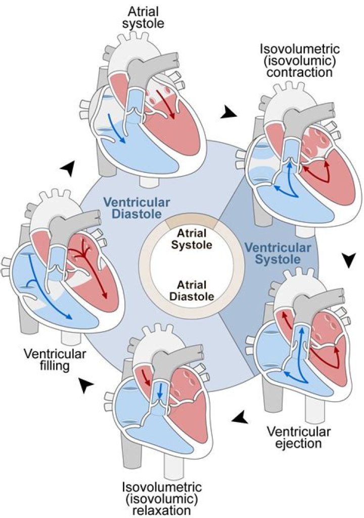
During a single cardiac cycle, the atria and ventricles do not beat simultaneously; the atrial contraction occurs prior to ventricular contraction. This timing delay allows for proper filling of all four chambers of the heart.
What Wave does ventricular contraction occur?
QRS wave
The QRS wave of the electrocardiogram represents ventricular depolarization, which is followed by contraction and an increase in pressure in the ventricles (ventricular systole). The T wave of the ECG represents ventricular repolarization and relaxation of the ventricular muscles (ventricular diastole).
Which wave is associated with a ventricular systole?
QRS complex
The QRS complex refers to the combination of the Q, R, and S waves, and indicates ventricular depolarization and contraction (ventricular systole). The Q and S waves are downward waves while the R wave, an upward wave, is the most prominent feature of an ECG.
How is atrial and ventricular contraction controlled?
This depolarisation and contraction of the heart is controlled by a specialised group of cells localised in the sino-atrial node in the right atrium- the pacemaker cells. These cells generate a rhythmical depolarisation, which then spreads out over the atria to the atrio-ventricular node.
Is atrial depolarization the same as contraction?
Atrial depolarization initiates contraction of the atrial musculature. As the atria contract, the pressure within the atrial chambers increases, which forces more blood flow across the open atrioventricular (AV) valves, leading to a rapid flow of blood into the ventricles.
How does isovolumetric contraction occur?
In cardiac physiology, isovolumetric contraction is an event occurring in early systole during which the ventricles contract with no corresponding volume change (isovolumetrically). This short-lasting portion of the cardiac cycle takes place while all heart valves are closed.
Is depolarization contraction or relaxation?
When the electrical signal of a depolarization reaches the contractile cells, they contract. When the repolarization signal reaches the myocardial cells, they relax. Thus, the electrical signals cause the mechanical pumping action of the heart.
Is systole a contraction?
Systole, period of contraction of the ventricles of the heart that occurs between the first and second heart sounds of the cardiac cycle (the sequence of events in a single heart beat). Systole causes the ejection of blood into the aorta and pulmonary trunk.
Is systole a contraction or relaxation?
Systole is the contraction phase of the cardiac cycle, and diastole is the relaxation phase. At a normal heart rate, one cardiac cycle lasts for 0.8 second.
Is depolarization systole or diastole?
Initially, both the atria and ventricles are relaxed (diastole). The P wave represents depolarization of the atria and is followed by atrial contraction (systole).
When does the atrial contraction occur at high heart rates?
At high heart rates when there is less time for passive ventricular filling, the atrial contraction may account for up to 40% of ventricular filling. This is sometimes referred to as the “atrial kick.”
How are ECG waves related to atrial and ventricular systole?
The ECG waves predict the timing of atrial and ventricular systole and diastole. At a heart rate of 75 beats per minute, the timing is as follows
What happens to the left ventricle during atrial contraction?
Atrial contraction normally accounts for about 10% of left ventricular filling when a person is at rest because most of ventricular filling occurs prior to atrial contraction as blood passively flows from the pulmonary veins, into the left atrium, then into the left ventricle through the open mitral valve.
How is the contraction of the atrial chambers initiated?
It is initiated by the P wave of the electrocardiogram (ECG), which represents electrical depolarization of the atria. Atrial depolarization initiates contraction of the atrial musculature. As the atria contract, the pressure within the atrial chambers increases, which forces more blood flow across…